
The risk of bone fractures is an emerging medical, social, and economic threat characterized by a systemic impairment of bone mass, strength, and fine structure, which increases the susceptibility of fragility fractures. This risk is caused by an imbalance in the physiological and cellular turnover of the bones in the bone by endocrine factors and / or autoclaving / paracrine (Fig.1.2b), which adversely affects peak bone mass and / or structural balance. A patient with osteoporosis has a greater tendency to fractures of the spine and femur, even if other bones may also be affected.
Osteoporotic fractures of the hip and spine lead to an increased mortality rate and are associated with important medical complications, such as pneumonia or thromboembolic disease caused by chronic paralysis that has a negative economic impact on public health, which is a global public health concern and produces a socio-economic impact. Substantial burden, which deserves to be addressed in an evidencebased and cost-effective manner, considering many risk factors. Given these initial considerations, treating osteoporosis is an important area of study where most efforts converge. Treatment should prevent vertebral fractures (which mostly depend on the density of the trabecular bone and structure) and nonvertebral fractures (which depend mostly on the thickness of the shell and the porosity). This can be accomplished by inhibiting bone resorption and / or by stimulating bone formation.
Bone remodeling or modeling activity varies between cortical and trabecular bone sites, and this difference can often explain the relative lack of efficacy of anti-absorption drugs on nonvertebral fractures since their effect is higher on the trabecular bone than cortical bone.
At the same time, it is evident that the engineering mechanics-based assessment of fracture risk, as it is the basis of structural design in civil and mechanical engineering, must have the ability to overcome the deficiencies based on the purely statistical and population related approaches referred to previously. Accordingly, a finite element risk assessment based on computed tomography of skeletons remains a central wish in bone biomechanics. This topic has been dealt with widely, but broad clinical application has not yet been achieved. Effective confrontation of these unfavorable trends requires effective identification of high-risk individuals, which is a sine qua non for effective primary prevention.
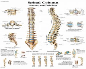
Figure 1.1 - Human spine anatomy and pathology
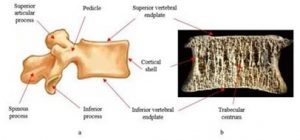
Figure 1.2 – a schematic view and b - actual image of the body of the transected lumbar spine in the mid-sagittal plane, showing the cortical layer
In modern conditions, when visualizing the area of a pathological fracture, a combination of technical and informational means is used. There are different methods for determining osteoporosis in a person and among them they are compared here to provide the best diagnostic suggestions for an accurate estimation of osteoporosis. The information base for pathology diagnosis is based on the creation of special quantitative diagnostic techniques, which can be based on a comprehensive physical and mathematical analysis of the interaction of the bones under study with radiation. For this purpose, mathematical models of these bones are used and their interaction with radiation is described. The most common technical means for radiological diagnosis of pathological bone fractures are X-ray diagnostic devices such as X-ray duplexing.
X-ray machines allow X-rays to be received, which determine the most information about the internal structure of the bone, and which can be used later in the diagnosis of fractures. In recent years, digital radiography has found an application, with special panels being the future of x-rays. After exposure to x-rays, a latent image of the object remains on it. When the panels are examined with a laser beam, the energy is released in the form of a glow, the intensity of which is proportional to the absorbed X-ray dose. This glow is recorded by the image detector and is digitized.
The resulting image can be displayed on the screen, printed on the printer, and stored in the computer's memory. X-ray bone measurement is a unique technology that basically allows us to operate osteometrics to identify the smallest changes in bone tissue and to make measurements with high accuracy for a minimum scan time (20 seconds) at a low dose rate. All X-ray-based methods provide a measure of bone mineral density (BMD), but other structural aspects of bone have been demonstrated to be important in determining fracture risk, such as mechanical features and elastic properties, which cannot be assessed using density scale techniques. Among the techniques most used, dual Xray absorptiometry (DXA) is the current 'gold standard' for diagnosing osteoporosis and predicting fracture risk.
Unfortunately, as other X-ray based technologies, DXA has specific limitations (eg, use of ionizing radiation, large size of equipment, high cost, limited availability) that hinder its application for population screening and primary care diagnostics. Dual-energy X-ray absorptiometry (DEXA). The measuring area is the spine, hip, or overall body. DEXA is the most popular method for measuring bone density.
BMD with DEXA is painless and does not require injections, invasive procedures, anesthesia, special diet, or any other pre-preparation. During a DEXA test, the patient lies fully on a padded table while the system examines one or more areas of bone (usually the lower part of the spine or hip). The entire test usually takes only a few minutes to complete.
Table 1 - Comparative characteristics of bone and joint imaging methods
|
Med - image devices |
Tubular Bones | Spine | Soft formations
& joints |
| X-ray | ++++ | ++++ | +++ |
| Ultrasound | ++ | ++ | |
| X-ray CT | +++ | ++++ | ++ |
| MRI | + | ++++ | ++++ |
| Radioisotope Diagnostics | + | + | ++ |
Conclusions & Recommendations The task of improving the methodological and technical support for the diagnosis of fractures in a large-scale population survey is important, despite significant progress in developing new tools to study the human bone system. When creating new quantitative methods of diagnosing fractures, it is necessary to develop the capabilities of traditional Xray diagnostics, supplement them with the latest methods and tools to obtain and process diagnostic information, Which should be based on the results of the physical and mathematical studies. Technical and informational tools for fracture diagnosis should provide an opportunity to conduct a mass survey of the population at the lowest material cost to retool existing diagnostic complexes. To develop an effective automatic system to detect and recognize bone fractures, a clear understanding of the human skeletal system and fractures is required.