

The air passageway as well as an appropriate contact of the air flux with the mucous membrane is important. Under normal conditions the healthy nasal anatomy forms special aerodynamic elements that control the air flux by way that all respiratory functions can be fulfilled adequately [1]. It is thus interesting to study the most important aerodynamic processes occurring in the nasal airway specifically with breathing disorders, and to identify the most important respiratory functions of the upper respiratory system [2].
The curvature of the nasal septum is the deviation of the septum into both or one side of the midline (see Fig. 1, b, e, h). It is manifested by difficulty or lack of nasal breathing through one or both halves of the nose. Minor curvatures of the nasal septum, which do not cause any symptoms, occur in more than 90% of the population.
The main types of curvature of the nasal septum are:
- Physiological - is associated with heredity or is a consequence of improper development of the facial skeleton due to a discrepancy in the growth rate of cartilage and bone tissue;
- Compensatory - result of influence of any irritating factors, for example, polyps, foreign bodies;
- Traumatic - the result of a dislocation or fracture of the nose.
Chronic rhinosinusitis is a long (at least 3-4 weeks) inflammation of the mucous membrane of the nasal cavity, passing to the nasal mucosa and causing severe edema of the tissues (see Fig 1, c, f and i). As shown in Fig 1, a), d), g) - schematic illustration and computer tomograms in the frontal and axial projections are respectively normal; b), e), h) is a schematic illustration and computer tomograms in the frontal and axial projections, respectively, with the distortion of the nasal septum to the right; c), f), i) - schematic illustration and computer tomograms in the frontal and axial projections, respectively, in chronic rhinosinusitis (Cx - middle line, SR, SL - denotes the plan of the right and left nasal passages respectively).
а) b) c)
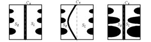
d) e) f)
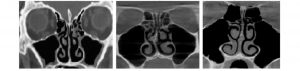
g) h) i)
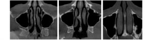
Figure 1 – Nasal cavity, normal and with typical chronic diseases
Changes in the architectonics of the nasal cavity led to a disruption in the aerodynamics of the nose, making it difficult to remove secretions from the paranasal sinuses, which leads to an increased propensity to develop inflammatory and allergic diseases of the upper respiratory tract.
The results will prove demonstrate that the intimate relation between nasal form and flow can be explored in greater detail than hitherto possible.By outlining means to compare complex airway geometries and demonstrating the effects of rational geometric simplification on the flow structure, this work offers a fresh approach to studies of how natural conduits guide and control flow. The concepts and tools address issues that are thus generic to flow studies in other physiological systems.
The idea was to examine breathing of groups of people from different regions according to the structures of their skull (Fig. 2), there is different climate, different biological race, so it would be possible to detect some another in physiology of nose breathing. Also, possible to overlap anatomical points of skull to CT scan for recognizing race.

Figure 2-Anatomical Structure of the facial skull
Skeletal indicators of race, focus primarily on top skull structure. We compare the skulls structure and can measure the distance between some points by using the ruler tool of CT viewer (Fig. 3).
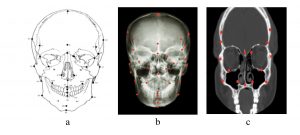
Figure 3 – Skull landmarks:
a -sketch of facial skull with land marks; b - X-ray image; c - CT normal slice
CT Signs help to understand the differences parameters, thicknesses and volumes for different race according to the structure of their skull. The most important signs are: Lower eye border, nasal index, nasal cavity shape, nasal bones, nasal overgrowth, nasal sill or dam, lower nasal spine. CT Signs can solve the task to find specific signs for definition of race easier.
These differences are important factors to know and to understand the dynamic of air flow, by measuring the different points that help to understand the different alterations in passage size, different structural composition and thickness of each race such as cranial index, nasal cavity and nasal bone.
These new results make use of improvements to computational and experimental techniques and resources, which now allow key dynamical features to be investigated, and offer rational procedures to relate variations in anatomical form.
References: